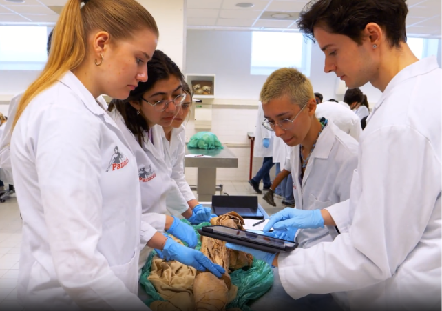
3D anatomy learning is rapidly evolving. In the dissection rooms of several University Medical Centers (UMCs) in the Netherlands, a quiet technological transformation is taking place. Where anatomical education has traditionally relied on textbooks, static atlases, and in-person dissections, a new workflow now captures human anatomical specimens as highly detailed digital twins.
At the heart of this process is photogrammetry — a technique that reconstructs three-dimensional geometry from hundreds or even thousands of overlapping photographs. Combined with automation, high-resolution imaging, and advanced post-processing, this method allows real human specimens to be preserved as immersive, interactive 3D models.
This is the foundation of Enatom’s reality-based E-learning platform.
Photogrammetry for anatomy
Photogrammetry works by identifying common visual features across many photographs taken from different angles. Specialized software analyzes these overlapping images and calculates the spatial position of each point in three-dimensional space. The result is a dense, accurate 3D reconstruction with true photographic texture.
To capture a single human anatomical specimen, hundreds of photographs are required. In practice, the number ranges between 600 and 3000 images, each taken at a resolution of 60 megapixels. The high resolution is crucial: it preserves fine anatomical details such as vascular branching, fascial layers, and subtle color differences between tissues.
However, high-quality photogrammetry is not simply about taking many pictures. It demands:
- Precise and consistent camera positioning
- Even coverage of the entire surface
- Stable lighting conditions
- Repeatable capture workflows
This level of precision is nearly impossible to achieve manually for complex anatomical structures. That is why automation became essential.
Image above: The process of photogrammetry involves taking hundreds of pictures of an object and create a point cloud from those pictures that can later be converted into a 3D textured mesh.
How Enatom Creates 3D Anatomical Models
The Enatom Scan Robot: Automation for Consistency and Precision
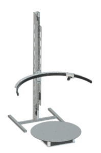 To ensure consistent placement of the camera and complete coverage of each specimen, Enatom developed a dedicated scan robot (called the ENAscanR) specifically designed for use inside hospital dissection rooms.
Enatom designs and builds its own automated systems to carry out photogrammetry with care and precision. From the earliest concept sketches to the final calibration, every stage of development is shaped in-house. This integrated approach allows Enatom to refine each detail, ensuring accuracy, consistency, and performance. By keeping design, engineering, and production under one roof, we maintain full control over quality and innovation. At Enatom, delivering high-resolution digital reality is the goal.
Image on the left: CAD design of the ENSscanR.
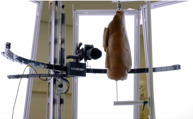 Image above: Scanning with the ENAscanR – in the vertical configuration
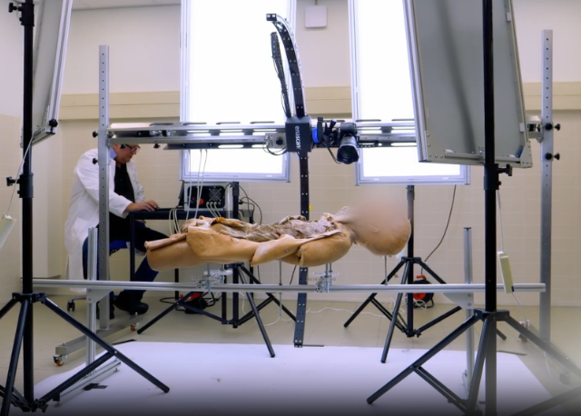 Image above: Scanning with the ENAscanR – in the horizontal configuration
This robotic system guarantees:
- Controlled camera trajectories
- Repeatable capture sequences
- Uniform overlap between images
- Stable geometry across large datasets
Depending on the specimen, either the camera rotates around the preparation, or the specimen itself rotates while the camera remains fixed. Both approaches are optimized to ensure complete surface coverage without introducing distortion or parallax inconsistencies.
The scanning process takes place in the dissection rooms of various Dutch UMCs that collaborate with the project. This is made possible by the well-organized and ethically structured body donation programs in the Netherlands. These programs provide medical education and research with access to human anatomical material, and they form the essential foundation for creating realistic digital models.
The work is conducted with respect, professionalism, and in close cooperation with the participating institutions.
Preparing the Specimen: Custom Rigs for Each Anatomy
Before scanning can begin, each specimen must be carefully mounted. No two anatomical preparations are identical — size, weight, fragility, and orientation vary greatly.
For this reason, a custom rig (support structure) is designed and built on location for each specimen. The rig is tailored to:
- Securely suspend or fix the specimen
- Minimize visual obstruction
- Allow full 360-degree coverage
- Maintain anatomical orientation
The goal is simple: provide maximum stability while keeping the support structures as invisible as possible during scanning.
Once mounted, the automated capture begins. Over the course of the session, between 600 and 3000 high-resolution photographs are taken, documenting every visible surface of the specimen.
From Images to 3D Model: RealityScan Processing
After acquisition, the photographs are processed using photogrammetry software — specifically RealityScan. The software aligns all images, reconstructs a dense point cloud, generates a polygonal mesh, and projects the original photographic textures onto the 3D surface.
This computational stage transforms thousands of flat images into a cohesive, navigable, high-resolution 3D model.
The output at this stage is already highly detailed, but it is not yet ready for educational use.
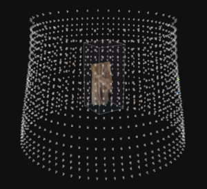
Image above: Alignment of hundreds of camera positions in Reality Scan
Post-Processing: Cleaning, Repairing, Refining
Because specimens are mounted in rigs, some structural elements inevitably appear in the raw reconstruction. These supports must be digitally removed.
Post-processing involves:
- Removing visible rigging hardware
- Repairing holes left behind after removal
- Reconstructing missing geometry
- Rebuilding realistic texture in edited areas
- Homogenizing mesh density
This step requires both anatomical knowledge and digital sculpting- and texturing expertise. The goal is not to beautify or idealize the anatomy, but to preserve authenticity while ensuring technical usability.
Every correction is performed carefully to maintain anatomical integrity.
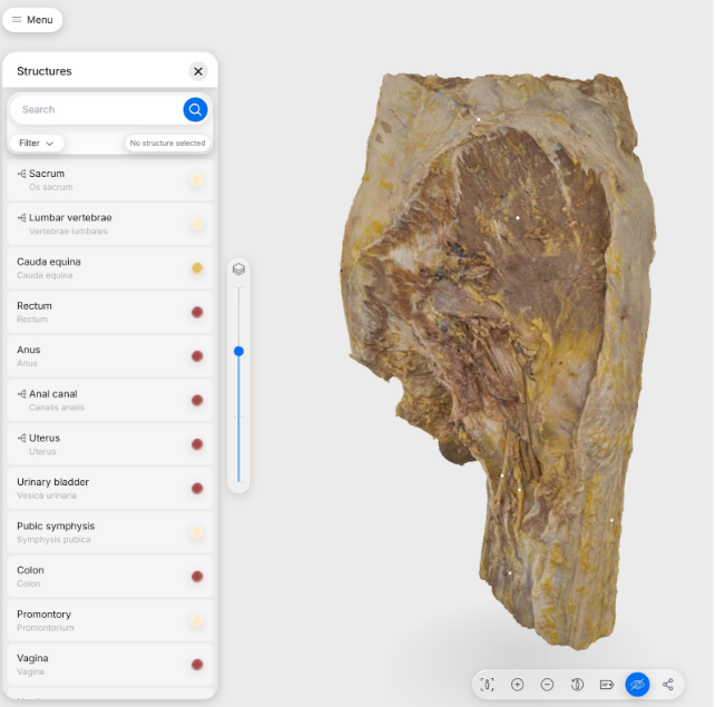 Image on the left:
The resulting highly detailed 3D mesh model.
A Proprietary File Format for Performance and Accessibility
Once cleaned and optimized, the 3D models are converted into a special file format developed by Enatom. This format is designed to:
- Stream efficiently over the internet
- Maintain high visual fidelity
- Perform smoothly on different hardware
- Support interactive anatomical exploration
The result is a lightweight yet detailed digital specimen in point cloud format, that can be accessed anywhere.
Enatom: Platform-independent highly realistic 3D anatomical models.
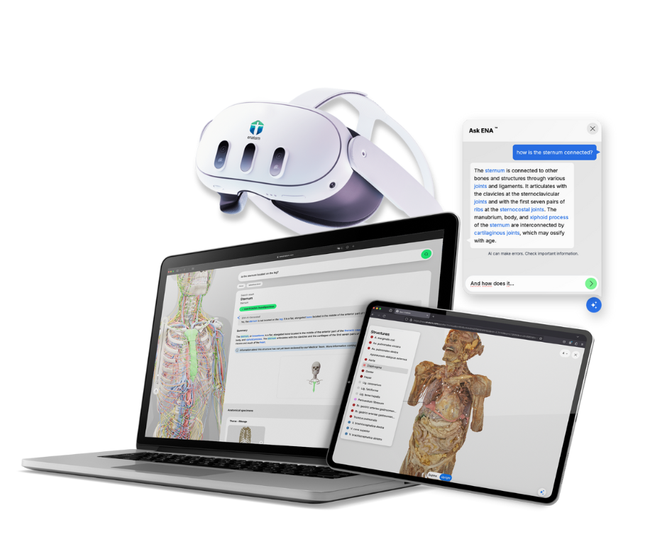
Enatom has developed a platform-independent application for viewing these highly realistic 3D anatomical models. The app runs on any device with an internet connection — desktop, tablet, laptop, or even mobile.
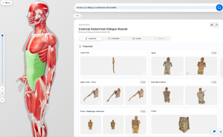
Image above: The user interface of the Enatom app offering many scanned specimen, AI search, 2D text book images and explanatory information with many cross links.
The Enatom app is more than a viewer.
The application includes:
- AI-powered search functionality
- Extensive textual anatomical information
- Structured knowledge layers
- Explanations of relationships between anatomical structures
Users can explore not only what a structure looks like, but also how it connects functionally and spatially to neighboring anatomy.
VR and AR: A Virtual Dissection Room
The app also supports VR (Virtual Reality) and AR (Augmented Reality) devices. In VR, users experience a fully immersive environment — a digital dissection room where real human specimens can be examined from any angle.
In AR, models can be projected into the user’s physical space, allowing anatomy to appear directly on a desk or in a classroom.
This creates a powerful educational experience: a realistic, spatial, embodied way of learning anatomy without replacing traditional dissection, but extending its accessibility.
Anatomical Diversity: Beyond the Textbook
One of the most important aspects of this project lies in its philosophical foundation.
Textbooks typically show idealized anatomy — clean, symmetrical, simplified. But real human bodies are diverse. Vascular branching varies. Muscular insertions differ. Pathologies alter structures. No two bodies are identical.
Enatom has many more specimens scheduled for scanning and inclusion in the platform. Each new model adds to a growing digital archive of anatomical diversity.
This diversity is precisely what traditional media struggle to represent. Static images cannot convey the full range of natural variation. Even cadaver labs provide limited exposure to variation within a small group of specimens.
By contrast, a scalable digital archive can preserve and present many unique anatomical realities side by side.
The Future of Anatomy Education
Enatom represents a new phase in anatomical education: reality-based E-learning V2.0.
It does not replace cadaveric dissection.
It does not replace textbooks.
Instead, it extends them.
By combining:
- High-resolution photogrammetry
- Automated robotic capture
- Advanced 3D reconstruction
- Intelligent software design
- VR and AR immersion
Enatom bridges physical anatomy and digital accessibility.
The dissection room remains the origin. The donor remains central. The technology serves as a translator — preserving real human anatomy in a form that can educate globally, repeatedly, and interactively.
Every scan is unique. Every body tells a different anatomical story.
And that diversity — preserved through precision scanning and presented through immersive technology — is exactly what defines the future of anatomy education.