
When people first see Enatom’s realistic 3D anatomy models, they often assume the work mainly happens behind a computer screen. They imagine artificial intelligence, advanced rendering software, or 3D artists digitally building the human body from scratch.
In reality, accurate anatomy learning starts somewhere else entirely: the dissection room.
At Enatom, every anatomical 3D model begins with real human anatomy. Together with leading medical universities, we work with authentic anatomical specimens prepared for medical education and scientific research. These are not artistic interpretations or simplified textbook drawings. They are real anatomical preparations, created and dissected by experienced anatomists who understand exactly which structures students need to study.
That difference matters.
Why realistic anatomy models matter in medical education
Medical students need more than clean illustrations. They need to understand the true complexity of the human body: spatial relationships, anatomical variation, tissue depth, and how structures exist in relation to one another.
Traditional anatomy atlases and 2D diagrams remain valuable, but they often cannot fully prepare students for what they encounter in clinical practice or the dissection lab. Realistic 3D anatomy models help bridge that gap.
By studying real anatomy in 3D, students can better understand:
- Anatomical depth and orientation
- Relationships between muscles, nerves, vessels, and organs
- Natural anatomical variation
- Complex regions such as the pelvic floor, skull base, and thorax
- Clinical anatomy in a more realistic context
This is one of the reasons why 3D anatomy learning platforms are becoming increasingly important in modern medical education.
Building anatomical 3D models starts with high-quality source material
Before we can create interactive anatomy learning modules or photorealistic anatomy models, we first need exceptional anatomical source material.
A digital anatomy model is only as good as the anatomical preparation behind it.
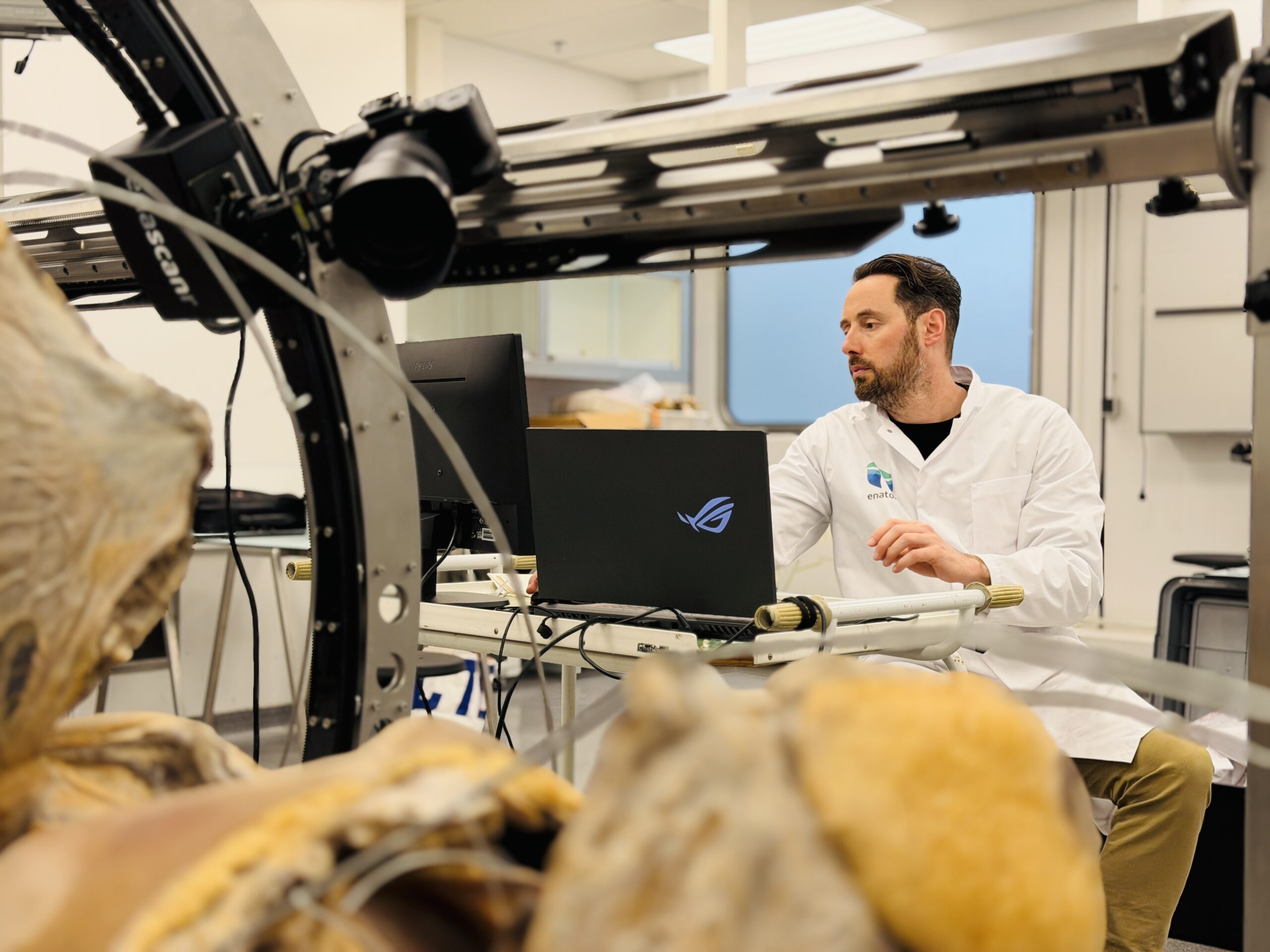
Together with my colleague Huib Schippers, I have spent hundreds of days inside dissection rooms documenting anatomical specimens for our growing library of realistic anatomy models.
This hands-on process is essential. By working directly alongside anatomists and universities, we can capture real human anatomy with the precision required for reliable medical education.
Every specimen is photographed from multiple angles in extremely high resolution. Small details matter:
- Tiny nerve branches
- Vascular pathways
- Tissue layers
- Organ relationships
- Surface textures
- Anatomical depth
Lighting consistency, camera positioning, resolution, and preparation quality all directly influence the final model.
This is not commercial product photography. It is scientific anatomical documentation.
From dissection room to photorealistic 3D anatomy model
Once the anatomical preparation has been documented, the digital reconstruction process begins.
Using advanced photogrammetry and visualization techniques, we transform real anatomical specimens into interactive, photorealistic 3D anatomy models that students can explore on laptops, tablets, phones, and VR devices.
I have worked in visualization since 2004, and throughout that time I have seen how digital technology can either oversimplify reality or help people understand it more clearly.
At Enatom, we focus on realism.
We do not aim to create anatomy that looks artificially clean or visually “perfect.” We want students to study real anatomy as it actually appears in the human body — including real textures, natural complexity, and authentic spatial relationships.
That realism is what makes digital anatomy learning more effective.
Students can:
- Rotate structures freely
- Zoom into difficult anatomical regions
- Study layers step-by-step
- Repeat complex material without lab limitations
- Learn anatomy outside scheduled dissection hours
- Better prepare for practical exams and clinical training
Expanding access to anatomy education worldwide
Not every medical student has access to a dissection room.
In many countries, anatomy education is limited by:
- High infrastructure costs
- Limited specimen availability
- Regulations around body donation
- Short laboratory access times
- Growing student populations
Even universities with strong anatomy programs often face limitations in capacity and teaching time.
Digital anatomy learning helps solve part of this problem.
With realistic anatomical 3D models, students can study human anatomy from anywhere while universities can extend anatomy teaching beyond the physical lab.
This does not replace traditional anatomy education. It strengthens it.
Our goal at Enatom is straightforward: help more medical students worldwide gain better access to high-quality anatomy learning based on real human anatomy.
Collaboration with universities is essential
A large part of my work involves maintaining long-term collaborations with medical universities and anatomy departments.
These partnerships are the foundation of everything we create.
We collaborate with institutions including:
- Amsterdam UMC
- UMC Groningen
- Maastricht UMC+
- Erasmus MC
These universities contribute anatomical expertise, educational validation, and carefully prepared specimens. We contribute imaging, visualization, and interactive 3D technology.
Together, we create anatomy learning tools that combine scientific accuracy with digital accessibility.
The future of realistic anatomy learning
As medical education evolves, students increasingly expect flexible, interactive, and accessible ways to study anatomy.
But innovation should never come at the cost of anatomical accuracy.
The future of anatomy learning is not about replacing real anatomy with digital tools. It is about preserving real anatomy digitally and making it accessible to more students worldwide.
That is why everything still starts in the dissection room.
Not with software.
Not with rendering.
Not with animation.
But with real human anatomy, prepared by experts, documented with precision, and transformed into realistic 3D anatomy models designed for medical education.
Because if you want to build the most realistic 3D human anatomy model possible, you first need the real human body.
